Dr. Rob Dillon, Coordinator


Thursday, December 15, 2016

Freshwater gastropods take to the air, 1991

Editor’s Note – This essay was subsequently published as: Dillon, R.T., Jr. (2019d) Freshwater Gastropods Take To The Air, 1991.  Pp 95 - 100 in The Freshwater Gastropods of North America Volume 4, Essays on Ecology and Biogeography.  FWGNA Press, Charleston.

Thirty years ago, when I signed my contract with Cambridge University Press to deliver the book that ultimately became The Ecology of Freshwater Molluscs [1] I had a larger project in mind.  The original title of the book was to have been, The Evolutionary Ecology of Freshwater Mollusca, and I planned to cover population genetics all the way up to speciation, as well as life history, competition, predation, communities, and the more traditionally ecological topics.  That was too much.

But before I realized that I had bitten off more than I could chew, I spent the summer of 1991 working on a chapter about freshwater mollusk gene flow.  It included subsections on straight-ahead crawling, drift, and “phoresy” under which I lumped all the cases where mollusks are carried passively by anything, including fish carrying the glochidia of freshwater mussels.  I even covered human-mediated invasions in that chapter, or at least tried.  Way, way too much!

I never submitted my gene flow chapter for publication.  But I rediscovered a hard copy again as I was moving out of my old office this past August, together with a bunch of raw data and tables and figures, and analyses on tractor-feed printer paper, and it re-awakened a dormant interest [2], and gave it a fresh slant.  How much has science advanced in 25 years?  The quick answer is that in some areas, progress has been Yuuuuge!  But in other areas, not so much. 

So for the next couple months we’ll focus on dispersal in freshwater gastropods.  Any study of which, in 1991 or in the present day, might well begin with the charming 1965 “presidential address” offered by W. J. Rees, “The aerial dispersal of Mollusca” [3].  Rees accumulated dozens of published references, notes, stories and anecdotes about both bivalves and gastropods, both terrestrial and aquatic, both pinched onto the feet and riding upon the shoulders of birds, bugs and bats, and even occasionally sucked up in cyclones and spat out on the bowlers of unsuspecting Englishmen. 

Rees collected 10 reports of freshwater limpets on aquatic insects, and I found two additional cases published between 1965 and 1991 – van Regteren Altena (1968) reporting 6 ancylids on the elytra of an aquatic beetle in Surinam, and Rosewater (1970) reporting 2 Laevapex taken from the elytra of a dytiscid beetle captured in a Florida light trap [4].  Does that total surprise you as much as it surprises me?  Even I, who have devoted my entire professional life to malacology, think of freshwater limpets as among the most obscure of all God’s creatures.  They’re just tiny little brown bumps, for Heaven sake!  Would you have guessed 12 published reports of freshwater limpets on flying water bugs?  That’s probably more than the total number of papers published on all other aspects of ancylid biology in North America combined.

The overland dispersal of non-limpet families of freshwater gastropods seems to be significantly less common.  Rees conveyed the report of a single Australian worker, who on different occasions found 6 species of snails on the feet of ducks.  I caught one more – the report of Roscoe (1955) of juvenile Physa, Lymnaea, and Helisoma on the feathers of a white ibis collected in Utah [5].

The review of Rees was followed by a couple really cute experimental papers published between 1965 and 1991 – those of Boag [6] on floating feathers and Malone [7] on severed killdeer feet.

Malone simply shot a killdeer, mounted its legs on a pair of props, and moved them through shallow water where the birds had been observed feeding.  He inspected the legs frequently, never allowing them to remain stationary for more than three minutes.  Two resident snails, Lymnaea humilis (“obrussa”) and Promenetus exacuous [8] often attached passively, drawn by the surface film or dislodged from vegetation as the feet moved through.  Although adult Lymnaea clung no more than five minutes if the feet moved, Promenetus and juvenile Lymnaea remained attached indefinitely.  All snails fell off when re-immersed in water.  Malone reported that L. humilis could survive 2 – 14 hours out of water, and Promenetus 5 – 14 hours, although admittedly not in conditions designed to duplicate flight.


Malone also reported that mature Lymnaea attached to the feathers of a (presumably footless) killdeer body left floating in shallow water for only three minutes.  Continuing along these lines, Boag [6] reared populations of Lymnaea stagnalis, L. elodes, and Helisoma trivolvis from eggs to age three months, periodically floating a feather in each culture pail.  Feathers with adhering snails were removed and placed in an air jet simulating flight speed for up to 15 minutes.  He record the sizes of the snails attached, their detachment rates, and their subsequent viability.  All of the (many) hundred snails found attached to feathers were in the 1 – 3 mm size range.  Although Boag did not monitor the size distributions of the base populations, it seems certain that the volunteer aviators tended to be significantly smaller, especially as the months of his experiment advanced.  The figure below shows his results combined over the duration of the experiment for the population with the largest data set, L. elodes.

Although riding on a feather must at best be only a rough approximation of riding on a bird, it is safe to conclude that either experience is quite rigorous.  Factoring both attachment and survivorship together, Boag’s data show that 56% of the L. elodes would arrive viable after 1 minute of flight, 17% after 5 minutes, 16% after 10 minutes, and only 4% after 15 minutes.  Figures were comparable for L. stagnalis and suggested an even lower probability of successful colonization for H. trivolvis.
Juvenile L. elodes on feathers exposed to simulated flight [6]
Three points emerge from the consideration of Malone’s data together with those of Boag.  First, it is clear that the attachment of snails to birds is the easy part, although perhaps only the smallest are likely to hold on.  And survivorship of these (typically small) individuals riding on birds is probably nowhere near the hours suggested by Malone, but more like minutes.  But finally, since the absolute frequency at which snails become attached to birds may be surprisingly high, even a 4% survival rate may be sufficient to render aerial dispersal a significant factor in initial colonization and subsequent gene flow among populations of freshwater snails.

In 1991 I also reviewed a second set of experiments published by Malone [9] assessing the likelihood of freshwater gastropod dispersal via gut transport.  It’s not great.  Malone fed caged ducks large volumes of aquatic vegetation, to which were attached the adults, juveniles, and egg masses of Physa and Helisoma.  Although he did not offer an estimate of the number of individual snails ingested, one can indirectly infer that this was surely in the thousands.  While no adults or juveniles were identified in the feces, Malone recovered 9 viable Physa embryos.  He also tried feeding about 500 – 1000 Physa egg masses and 200 – 400 Helisoma egg masses to killdeer, collecting feces in aerated water.  A total of 17 viable embryos were recovered.

Almost all of the material above was extracted from my 1991 chapter on dispersal.  So what progress have we made in studies on the aerial dispersal of freshwater gastropods since?  A fair amount, actually.  Tune in next month!


Notes

[1] Dillon, R. T. Jr.  (2000) The Ecology of Freshwater Molluscs.  Cambridge University Press. 509 pp.

[2] This is the second essay I have posted on the subject of aerial dispersal in freshwater gastropods, the first coming way back in November of 2005.  I also invoked a gene flow mechanism I termed the “sticky bird express” just this past April.  See:
  • Aerial Dispersal of Freshwater Gastropods [17Nov05]
  • Mitochondrial Superheterogeneity: What it means. [6Apr16]
[3] W. J. Rees (1965) The aerial dispersal of Mollusca.  Proc. Malac. Soc. Lond. 36: 269-282.

[4] van Regteren Altena, C. O.  (1968)  Transport of Ancylidae (Gastropoda) by a water beetle in Surinam.  Basteria 32:1.  Rosewater, J. (1970) Another record of insect dispersal of an ancylid snail.  Nautilus 83: 144-145.

[5] Roscoe , E. J. (1955)  Aquatic snails found attached to feathers of white-faced glossy ibis.  Wilson Bulletin 67: 66.

[6] Boag, D. A. (1986) Dispersal in pond snails: Potential role of waterfowl. Can. J. Zool. 64: 904-909.

[7] Malone, C. R. (1965) Killdeer (Charadrius vociferus) as a means of dispersal for aquatic gastropods. Ecology 46: 551-552.

[8] I’m a bit skeptical of this identification.  Perhaps Promenetus is significantly more common in NW Montana, where Malone conducted his research, than anyplace I have any personal observations.  But my guess would be Gyraulus parvus, not P. exacuous.

[9] Malone C.R. (1965) Dispersal of aquatic gastropods via the intestinal tract of water birds. Nautilus, 78: 135–139.

Monday, November 14, 2016

One Goodrich Missed: The skinny simplex of Maryville is Pleurocera gabbiana


Editor’s Note.  This is the third installment in a three-part series on the discovery of a cryptic pleurocerid species in East Tennessee.  You might want to back up and read my essays of 13Sept16 and 14Oct16 if you haven’t already.  A more technical version of the present essay was published in the FMCS Newsletter Ellipsaria 18(3): 10 – 12 [pdf] if you’re looking for something citable.

Longtime readers may remember the 2007 essay I wrote on Calvin Goodrich [1], who monographed the North American pleurocerids in a series of spare papers published by the University of Michigan 1939 – 1944.  Goodrich synonymized scores of specific pleurocerid nomina without rationale, and simply neglected to mention many more.  And by the end of his six-year effort, what had been a jungle of 1,000 species of North American pleurocerids [2] had been bush-hogged down to a merely untidy 150.  While admitting that Goodrich’s scholarship might not have been the most intellectually satisfying, here’s what I wrote in 2007: 
 “And Goodrich's review has proven to be of great use to malacologists working in American freshwaters today.  My 25 years of research on the population genetics of pleurocerids in the South suggests to me that the total number of biological species in this country will prove to be far less than 500, and indeed less than 100. I haven't found a biological species that Calvin Goodrich missed.”
 So back in September I reported the discovery of two species of Pleurocera cryptic under the common and widely-distributed P. simplex in East Tennessee, “fats” and “skinnies.”  And in October, I matched the fat simplex to the type population in Saltville, Virginia.  Now what are those “skinnies?”

My first thought was to refer back to Goodrich’s (1940) paper covering that part of the world [3], to see if one of the names Goodrich synonymized under P. simplex might be resurrected.  There were only five such names: warderiana, subsolida, densa, vanuxemii, and prestoniana [4].

Goodrich’s papers were unfigured, alas, and so my next move was to pull my trusty copy of Tryon (1873) off the shelves and try to picture-match [5].  If some of the shells of those putative simplex-synonyms figured in Tryon had been skinny, and if their original authors had been so kind as to suggest a type locality, I suppose I would have been strongly tempted to visit that locality and fetch a batch home for genetic work.  But none of the figures in Tryon corresponding to any of the names in Goodrich looked skinny in the least.

Figure 1.   Holotype of Goniobasis gabbiana (center) is compared to exemplars from S1 of Indian Creek (left) and S5 topotypic P. simplex (right).  The scale bar in mm.  A = Apex height, B = Body whorl height.

So I put the question aside for a while, and diverted my attention to other questions.  The thought of describing a new species did cross my mind. But good grief, number 1,001?  Does North America need another nominal species of pleurocerid?

In 2012 the focus of the FWGNA project shifted from East Tennessee to the Middle Atlantic States, and I started paying calls to all the national collections in the big cities of the east.  So it must have been the third or fourth day of my visit to the National Museum of Natural History on Constitution Avenue, and I got getting tired of logging drawers full of Physa.  And I thought I might just have a peek at the type collection, probably more out of intellectual curiosity than anything else.  Isaac Lea described over 400 species of pleurocerids [6] between 1831 and 1869, and (I suppose) a large fraction of his types are housed at the USNM, pretty much all together in a set of locked cabinets off to the side.  And by chance, my eyes happened to fall on USNM 118991, the lectotype of Goniobasis gabbiana.

Isaac Lea first published a brief Latinate description of Goniobasis gabbiana in his (1862) description of the new genus Goniobasis, followed by a more complete (English) description and figure in 1863 [7]. His locality data were uselessly vague: “Tennessee Prof. G. Troost; Alabama Prof. Tuomey.” The species nomen was passed along as valid by Tryon (1873) but was essentially forgotten by Goodrich, who listed it as a “species in doubt” in his 1930 Alabama paper [8] but neglected it entirely in his 1940 review of the Pleuroceridae of the Ohio River drainage system [3], which covered East Tennessee. Thus, although still valid, Goniobasis (or “Elimia”) gabbiana was not included in the more recent reviews of Burch (1989) and Turgeon et al. (1998).  In addition to the lectotype, the USNM holds just two lots labeled Goniobasis gabbiana, both from the nineteenth- century malacologist A. G. Wetherby, neither matching the type. To my knowledge, no pleurocerid lots are held under the nomen gabbiana in any other North American collection.

Figure 2.  The Goniobasis gabbiana collection at the USNM.

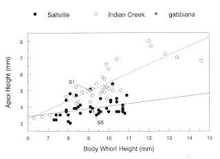
In Figure 3, the holotype of P. gabbiana is plotted by its apex height (A) and body whorl height (B) on that graph of Saltville fats and Indian Creek skinnies I developed last month [9]. The match in relative shell dimensions between USNM 118991 and Pleurocera population S1 of Indian Creek would appear nearly perfect. The evidence thus suggests that Pleurocera population S1 at Indian Creek and (by inference) skinny population S6s at Pistol Creek may be identified as Pleurocera gabbiana (Lea 1862), a nomen here revived after 150 years of obscurity.

The fat simplex /skinny simplex situation contrasts rather strikingly with most of my previous experience working with the systematics of the North American Pleuroceridae [10].  Typically intraspecific shell variation is so extreme that nineteenth-century taxonomists have recognized multiple nominal species, and even multiple genera, within single conspecific populations. Here’s the opposite situation, where interspecific shell variation is so slight that twentieth-century taxonomists have not distinguished any differences.

Figure 3. Shell apex (A) as a function of body whorl (B) in P. simplex topotypic population S5 and in population S1 from Indian Ck.  Exemplar shells are noted with arrows, cross marks the lectotype of Goniobasis gabbiana.

Our subsequent field surveys have uncovered 58 additional populations apparently referable to P. gabbiana -- often mixed with typical P. simplex -- locally abundant in small streams of the Tennessee River drainage from the vicinity of St. Paul, Virginia, southwest perhaps 100 km to Madisonville, Tennessee [11]. As we mount expeditions to catalog the rich biodiversity perhaps lying undiscovered in the most remote corners of the earth, we might profitably pause to examine the less exotic but equally remarkable biodiversity we have too often overlooked in our own backyards.


Notes

[1]  The Legacy of Calvin Goodrich  [23Jan07]

[2] Graf, D. L. (2001)  The cleansing of the Augean Stables, or a lexicon of the nominal species of the Pleuroceridae of recent North America, North of Mexico.  Walkerana 12: 1 – 124.

[3] Goodrich, C. 1940. The Pleuroceridae of the Ohio River system. Occasional Papers of the Museum of Zoology, University of Michigan 417:1-21.

[4] John Robinson and I added a sixth synonym in 2007, aterina (Lea 1863).  See:
  • Dillon, R. T., Jr. and J. D. Robinson. 2007. The Goniobasis ("Elimia") of southwest Virginia, I. Population genetic survey.  Report to the Virginia  Division  of  Game  and  Inland  Fisheries.  25 pp. [pdf]
[5] Tryon, G. W., Jr. 1873. Land and Freshwater Shells of North America. Part IV, Strepomatidae.  Smithsonian Miscellaneous Collections 253, 435 pp. Washington, D.C

[6] By my actual count, paging through Graf [2], Isaac Lea described 438 species of North American pleurocerids.  Not counting all the spelling errors and similar screw-ups by subsequent authors.

[7] Lea, I. (1862) Description of a new genus (Goniobasis) of the family Melanidae and eighty-two new species. Proceedings of the Academy of Natural Sciences, Philadelphia 14:262-272.  Lea, I. (1863) New Melanidae of the United States. Journal of the Academy of Natural Sciences, Philadelphia 5:217-356.

[8] Goodrich, C. 1930. Goniobases of the vicinity of Muscle Shoals. Occasional Papers of the Museum of Zoology, University of Michigan 209:1-25.

[9] The Fat Simplex of Maryville Matches Type [14Oct16]

[10] Papers documenting the situation where high levels of shell phenotypic variation have fooled previous authors into recognizing multiple species of pleurocerids where only one exists: Dillon, R. T., Jr. and J. D. Robinson (2011)  The opposite of speciation: Population genetics of Pleurocera (Gastropoda: Pleuroceridae) in central Georgia. American Malacological Bulletin 29:159- 168 [pdf].  Dillon, R. T., Jr.  2011.  Robust shell phenotype is a local response to stream size in the genus Pleurocera (Rafinesque 1818). Malacologia 53: 265-277 [pdf].  Dillon, R. T., Jr., S. J. Jacquemin and M. Pyron.  2013.   Cryptic phenotypic plasticity in populations of   the freshwater prosobranch snail, Pleurocera canaliculata.  Hydrobiologia 709:117-127 [pdf].  Dillon, R. T., Jr. 2014. Cryptic phenotypic plasticity in populations of the North American freshwater gastropod, Pleurocera semicarinata. Zoological Studies 53:31 [pdf].

[11] A detailed map comparing the distributions of P. simplex and P. gabbiana in East Tennessee and SW Virginia is downloadable from the FWGNA site.  [pdf map]

Friday, October 14, 2016

The Fat simplex of Maryville matches type


Editor’s Note.  This is the second installment of a three-part series on the discovery of a cryptic pleurocerid species in East Tennessee.  You might want to back up and read my essay of 13Sept16 if you haven’t already.  The present essay was published in the FMCS Newsletter Ellipsaria 18(2): 16 - 18 [pdf] if you’re looking for something citable.

So when last we left our intrepid malacologist, puzzling at his lab bench in the summer of 2008, he had come around to the realization that the population of Pleurocera simplex inhabiting Pistol Creek at the Courthouse Park in Maryville, Tennessee, was actually two reproductively isolated populations, fats (S6f) and skinnies (S6s).  Which population might be the bona fide P. simplex of Thomas Say (1825)?  And what might be the identity of the other?

I actually started out with a pretty good hunch.  In 2007 John Robinson and I ran allozyme gels on five populations of P. simplex from up in Virginia, including a nice sample from Say’s type locality in Saltville [1].  The Saltville population (S5) was fixed for the Oldh100 allele that turned out to be diagnostic for the Maryville S6f fats.  And interestingly enough, the simplex population John and I sampled from Indian Creek at Kesterson Mill way down at the southwest tip of Virginia (S1) was nearly fixed for that Oldh104 allele diagnostic for the Maryville S6s skinnies.  I hadn’t noticed any shell morphological differences between the Saltville simplex and the Kesterson Mill population at the time, however.

So in August of 2008 [2] I drove up to Virginia for additional samples from Saltville and from Kesterson Mill, which I returned to Charleston to run alongside a fresh batch of Maryville samples, to make sure all the bands matched up. Sure enough, the Maryville S6f fats were indeed genetically similar to P. simplex collected from its type locality at Saltville S5, not just at the Oldh locus but over all ten of the allozyme loci I have found informative for studies of this sort.  And the Maryville S6s skinnies were similar to Kesterson Mill S1.

Figure 1.  See footnote [3] for methodological details.

I also took the calipers to a bunch of the shells from Saltville and Kesterson Mill.  And sure enough, exactly the same pattern reappeared that we first noticed at Maryville.  Dividing the total shell length into body whorl height (B) and apex height (everything else, A), the (N = 37) shells I sampled from Kesterson Mill showed a significantly greater ratio of A to B than the (N = 40) shells from Saltville.  Figure 2 shows that the simple regression of apex height on body whorl height for the Saltville type population S5 was A = 0.157B + 2.46 (r = 0.36), very significantly different (ANCOVA t = -9.52, p < 0.0001) from the regression for the Kesterson Mill population S1, A = 0.556B - 0.09 (r = 0.84).  Saltville snails are fat, and Kesterson Mill snails are skinny.

Figure 2.

I have picked two shells directly off the regression lines to illustrate the difference between the two species.  So since the Maryville S6f fats match that lower-sloping S5 sample of N = 40 from Saltville both genetically and morphologically, and since Saltville is the type locality of Pleurocera simplex (Say 1825), clearly the Maryville S6f fats must be the bona fide simplex [4].

Then what might be the identity of the Maryville S6s skinnies, matching the higher-sloping S1 population from Kesterson Mill both genetically and morphologically? Tune in next time!


Notes

[1] Dillon, R. T., Jr. and J. D. Robinson. 2007. The Goniobasis ("Elimia") of southwest Virginia,  I. Population genetic survey.   Report to the Virginia Division of Game and Inland Fisheries.  25 pp. [pdf]

[2]  Yes, this was the same trip I featured last month, in which I resampled Maryville.  I’ve been sandbagging my story a little bit, to make it unfold in a more linear fashion.  To tell you the truth, I got the “hunch” mentioned in this month’s essay pretty much immediately upon reading my first Maryville simplex gel, and last month’s story unfolded pretty much simultaneously with this month’s story.

[3] Subsamples of 14 individuals from population S5 and 41 from population S1 were analyzed electrophoretically together with the 20f + 51s individuals from Pistol Creek I featured in last month’s blog post.  These data were combined with the data sets of N = 34 from population S5 and N = 37 from population S1 previously published by Dillon & Robinson (2007), and with the N = 17f + 13s data I had previously accumulated from Maryville. Then BIOSYS version 1.7 (Swofford & Selander, 1981) was used to calculate matrices of Nei's (1978) unbiased genetic identity (below the diagonal in Fig 1) and Cavalli-Sforza and Edwards (1967) chord distances between all pairs of control populations S1 and S5 and the two cryptic S6 populations co-occurring in Maryville. Chord distances were used as the basis for the neighbor-joining tree shown above the diagonal in Figure 1, as calculated using PHYLIP v3.65 program NEIGHBOR (Felsenstein, 2004).

[4]  Ultimately I only used that sample of 20 + 17 = 37 fats as my S6 Maryville control in Dillon 2011.  The existence of a second Pleurocera population at Maryville cryptic under simplex was not mentioned in my 2011 paper at all:
  • Dillon, R. T., Jr.  2011. Robust shell phenotype is a local response to stream size in the genus Pleurocera (Rafinesque 1818).   Malacologia 53:265-277. [pdf]

Tuesday, September 13, 2016

The Cryptic Pleurocera of Maryville


Editor's Note.  This is the first installment of a three-part series on the discovery of a cryptic pleurocerid species in East Tennessee.  The present essay was published in the FMCS Newsletter Ellipsaria 18(2): 15 - 16 [pdf] if you're looking for something citable.
"endless forms most beautiful and most wonderful have been, and are being, evolved."
I like to imagine that, as Charles Darwin wrote the poetic final clause of his masterwork, he may have been gazing over some well-curated systematic collection of molluscan shells [1].  Shells are captivating things, aren’t they?

This month’s installment in my long-running fascination with The Shell began in 2007, when I first glimpsed the phenomenon we ultimately christened “cryptic phenotypic plasticity" [2].  Using a survey of gene frequencies at a pair of highly polymorphic allozyme-encoding loci, John Robinson and I had just discovered that the pleurocerid populations we were identifying as Goniobasis acutocarinata, Goniobasis clavaeformis, and Pleurocera unciale on the basis of their strikingly different shell morphologies apparently constituted a single biological species [3].  Our samples had come from Indian Creek, a tributary of the Powell River on the Virginia/Tennessee line.  And I was curious to see if these intriguing results might extend generally through East Tennessee and into North Georgia, implying that the shell morphological distinction historically used to distinguish the pleurocerid genera Goniobasis/Elimia from Pleurocera might have no heritable basis.

So I picked three fresh study areas – the Little River drainage in the vicinity of Maryville (TN), the Conasauga drainage east of Etowah (TN), and the Coahulla near Dalton (GA).  And from each of these three regions I sampled three populations from the acutocarinata-clavaeformis-unciale-carinifera continuum to analyze together with my original data set from the Powell drainage.  And from each of these (now four) regions I also needed a control – a population of some common, widespread pleurocerid that everybody recognizes, the specific status of which nobody doubts.

Populations of Pleurocera simplex are very nearly omnipresent in small streams throughout Southwest Virginia, East Tennessee, and North Georgia.  They are also rather distinctive, with their dark, smooth, teardrop-shaped shells and strikingly black bodies.  Thomas Say described “Melania” simplex in 1825 from “a stream running from Abingdon to the Salt Works, and from the stream on which General Preston’s grist-mill is situated, as well as in a brook running through the salt water valley and discharging into the Holstein River.”  The eighteenth-century salt mines to which Say must have been referring are still identifiable in modern day Saltville (VA), and (indeed) snail populations matching Say’s small figure and description still inhabit streams in the vicinity.  And Say’s nomen “simplex” is among the oldest names available for any American pleurocerid [4], so is in no danger of being synonymized under anything else.

I included two populations of Pleurocera simplex (as “Goniobasis simplex”) in the first allozyme study I ever published, way back in 1980 [5], and added five fresh simplex populations to control that 2007 gray-literature report with John Robinson I referenced to open the present essay, extending west from the Saltville type locality across the Holston, Clinch and Powell drainages of SW Virginia [6].  Clearly Pleurocera simplex would be the perfect control for my follow-up study of the acutocarinata-clavaeformis-unciale-carinifera continuum now extending south, through East Tennessee and into North Georgia.  Am I right?

And so it was that on 7May08, I came to stand on the banks of Pistol Creek in Courthouse Park, Maryville.  What a lovely little city is Maryville, Tennessee!  Spreading like a cool oasis between the county courthouse and historic Maryville College I found a shady park full of happy picnickers and laughing children.  And Pistol Creek is chock full of Pleurocera clavaeformis acutocarinata, which was the focus of my attention on that sunny spring afternoon, mixed with a population of good old familiar Pleurocera simplex, the perfect control.  I was probably there no more than 30 minutes.  In my field notes I wrote, “Very pretty spot!  Friendly girls drove me off.”

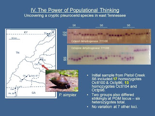
The genetic analysis would, of course, take somewhat longer.  I started running the gels for the study that was ultimately published as “Robust shell phenotype is a local response to stream size in the genus Pleurocera” [7] later that month, extending through the summer.  I pulled the first sample of 7 individuals from the bag labelled “Maryville simplex S6” on July 9, with another 15 individuals on July 11.  The PowerPoint slide below [8] shows photos of two of the nine gels I ran on 7/11/08 – one stained for octanol dehydrogenase (Octl), the other stained for octopine dehydrogenase (Octp).  The 15 Maryville simplex (labeled “S6”) were interleaved that day with ten simplex individuals from someplace else and six P. clavaeformis – don’t worry about those other 16 samples.


Much to my wondering eyes, the 15 “simplex” from Maryville S6 proved to comprise 9 homozygotes for Octl104 and Octp98, and 6 homozygotes for Octl100 and Octp96, with no heterozygotes in evidence at either locus.  There were also striking differences between the set of 9 and the set of 6 at the PGM locus.  I wrote in my lab notebook, “News Flash!!  There are TWO species inside Maryville S6.  Criminy!” 

Over the course of the next several runs, the sample of 30 snails I had collected from Maryville on the afternoon of 7May08 ultimately proved to include 17 of the one species and 13 of the other.  And almost immediately an important follow-up question began to nag me.  Might there have been some subtle morphological difference between these two sets of snails I had initially lumped together as Pleurocera simplex?  What about the shells?  Were they absolutely indistinguishable?  Alas, I had cracked the shells and thrown them all away when I froze my “Maryville simplex S6” sample back in May, following my standard practice.  

So in August of 2008 I returned to Maryville Courthouse Park for a second sample.  And on this second visit, I examined the shells much more critically.  Standing ankle-deep in Pistol Creek, I developed the impression that significant variation might exist in the simple shell proportions of the snails I had previously lumped together as P. simplex, particularly with respect to the relative heights of their body whorls.  Although the differences were slight – indeed negligible in juveniles and subadults – it seemed possible to me that the creek might be inhabited by an admixture of two snail populations with dark, smooth, teardrop-shaped shells and strikingly black bodies, one with “fat” shells and the other with “skinny.”

So, for the 71 Maryville snails I carried back to Charleston the next day, I measured the maximum shell dimension (or "shell height"), and body whorl height (B), defined as the length of the final 360⁰ of whorl, along the axis of coiling. I then defined apex height (A) as shell height minus body whorl height, and analyzed the relationship between body whorl height and apex height by analysis of covariance using the separate slopes model (JMP version 7).  I then classified my fresh sample of 71 individuals by their phenotype at 10 allozyme-encoding loci using standard methods.

A total of 20 snails proved homozygous for Oldh100, while 51 were homozygous for Oldh104, with no putative heterozygotes again in evidence. Differences were also very marked at the Opdh and PGM loci, although a few heterozygotes were observed in both groups.  The combined sample of 20 August snails plus 17 snails from the May sample showed Opdh96 = 0.946 and Pgm96 = 0.946, and the combined sample of 51 + 13 showed Opdh98 = 0.953 and Pgm102 = 0.852.  Again no variation was detected at the seven additional genetic loci examined.


The figure above compares the regressions of (A) on (B) for the two subsamples, the N = 20 fixed for Oldh100 and the N = 51 fixed for Oldh104.  Sure enough, the regressions are quite significantly different [9].  I provisionally designated the (N = 20) snails fixed for Oldh100 as population S6f, with f appended for “fat,”, and the (N = 51) snails fixed for Oldh104 as population S6s, for “skinny.”

One of these populations must surely match Thomas Say’s bona fide simplex from that “brook running through the salt water valley and discharging into the Holstein River,” I should hope!  But which one?  And what might be the identity of the other population?  Tune in next time!


Notes

[1] Charles Darwin, Freshwater Malacologist [25Feb09]

[2] I originally christened the phenomenon “Goodrichian Taxon Shift” in February of 2007, focusing entirely upon freshwater snails.  Stephen Jacquemin, Mark Pyron, and I broadened the concept into “cryptic phenotypic plasticity” in a (2013) paper we published in Hydrobiologia [PDF].  Most of the (now 17) blog posts filed under “Phenotypic Plasticity” in the blog index at right above touch upon the pervasive phenomenon of CPP in freshwater gastropod shells.  But see particularly:
  • Goodrichian Taxon Shift [20Feb07]
  • Pleurocera acuta is Pleurocera canaliculata [3June13]
[3] Dillon, R. T. & J. D. Robinson (2007b)  The Goniobasis ("Elimia") of southwest Virginia, II.  Shell morphological variation in G. clavaeformis.  Report to the Virginia Division of Game and Inland Fisheries.  12 pp.  [PDF]

[4] Thomas Say published four pleurocerid names in 1825: simplex, proxima, subglobosa, and fluvialis.  There are eight older pleurocerid names in the literature: virginica (Gmelin 1791), carinata (Brug. 1792), verrucosa (Say 1820), armigera (Say 1821), canaliculata (Say 1821), praerosa (Say 1821), catenaria (Say 1822), and carinifera (Lam. 1822).

[5] Dillon, R.T. and G.M. Davis (1980) The Goniobasis of southern Virginia and northwestern North Carolina: Genetic and shell morphometric relationships. Malacologia 20: 83-98. [PDF]

[6] Dillon, R. T. & J. D. Robinson (2007a)  The Goniobasis ("Elimia") of southwest Virginia, I.  Population genetic survey.  Report to the Virginia Division of Game and Inland Fisheries.  25 pp.  [PDF]

[7] Dillon, R. T. (2011)  Robust shell phenotype is a local response to stream size in the genus Pleurocera (Rafinesque 1818). Malacologia 53: 265-277. [PDF]  I featured these results in two blog posts:
  • Mobile Basin III: Pleurocera Puzzles  [12Oct09]
  • Goodbye Goniobasis, Farewell Elimia [23Mar11]
[8]  This slide comes from a presentation I made at the 2011 AMS meeting in Pittsburgh:
Dillon & Robinson (2011) When Cometh Our Reformation?  Molecular typology meets population genetics in the pleurocerid gastropod fauna of east Tennessee.

[9] The regressions of A = 0.34B + 1.96 (r = 0.69) for the Oldh100 group and A = 0.57B + 1.02 (r = 0.74) for the Oldh104 group differed significantly in their slopes, although not in their intercept. Separate-slopes ANCOVA returned a value of t = -7.14 (p < 0.0001) testing for a difference between the two groups, the group of 20 Oldh100 snails showing a very significantly lower apex (holding body whorl constant) than the group of 51 Oldh104 snails.

Thursday, August 18, 2016

The Classification of the Hydrobioids

Editor’s Note – This essay was subsequently published as: Dillon, R.T., Jr. (2019c) The classification of the hydrobioids.  Pp 205 - 210 in The Freshwater Gastropods of North America Volume 3, Essays on the Prosobranchs.  FWGNA Press, Charleston.

I confess that simply composing a header for the present blog post was a minor challenge for me.  How should I entitle an essay about a great big group of little tiny snails that really isn’t even a group?  The noun “hydrobioid” (without taxonomic status) has found widespread application in the literature [1], but if “tiny little odd-lot leftovers” could be Latinized into something that sounded fancy enough, that’s the word I’m fishing for.

The non-group of snails that will be the subject of this month’s blog post are the freshwater representatives of the Superfamily Rissooidea, vanilla gastropods bearing cusps around the base of their median radular tooth.  They give the impression of being smaller-bodied than is typical for the Class Gastropoda, although I’m not sure that’s true, and seem to include an unusually high proportion of shallow water, intertidal, or amphibious taxa.  Sexes are separate, as is true for most gastropods, the penis arising from behind the head.

And I should hasten to add the disclaimer that hydrobioids are not “leftovers” in the American West.  Throughout the Great Basin and Pacific drainages, hydrobioids can be the dominant (sometimes only) element of the freshwater gastropod fauna.  Their endemicity is legendary, and conservation concerns legitimate [2].  But here in The East, we naturally tend to focus on the pulmonates and the pleurocerids and the viviparids, and maybe, at the end of the afternoon, we might find a couple sullen little hydrobioids sucked onto a stick.

Through most of the 20th century, classifications of the hydrobioid taxa have typically recognized no more than five families worldwide.  Essentially all workers have always recognized the (huge, diverse) Hydrobiidae of Troschel (1857), to which Thiele (1929) added the Old World Micromelaniidae and the marine Rissoidae, for example.  Wenz (1939) also recognized three families: the Hydrobiidae, the Micromelaniidae, and the Truncatellidae, including Pomatiopsis.  Any of my readership with a taste for taxonomic arcana are referred to the 1993 monograph of Kabat and Hershler [3] for a tabulation of 16 different, and often strikingly discordant, 19th and 20th century hydrobioid classification systems.
Table 1 of Kabat & Hershler (1993)
The FWGNA project, at our birth in 1999, adopted the broad-sense definition of the Hydrobiidae that arose from Kabat & Hershler’s scholarly review.  So for the last 18 years we have recognized here in North American freshwaters the Bithyniidae (Gray 1857) with just one species, the Pomatiopsidae (Stimpson 1865) with just a couple species, and a huge, diverse Hydrobiidae with a zillion tiny little odd-lot leftovers.

But the dawn of molecular phylogeny was rendering Kabat & Hershler’s broad-sense concept of the Hydrobiidae obsolete even as we were adopting it.  In 1998 an international team of researchers headed up by Tom Wilke and George Davis began publishing the first gene trees with hydrobioid taxa at their tips [4].  The analysis of the Wilke team suggested that Troschel’s old family Hydrobiidae was polyphyletic, implying that many of the taxa previously considered subfamilies underneath the Hydrobiidae deserved promotion to the full family rank.

In retrospect, we might have updated our hydrobioid classification back there as early as 2001, and probably at least once or twice again in the mid-2000s as well.  I confess that I’ve just been letting the pot simmer.  But at some point one must serve.

So by 2013, the Wilke team, now including our good friends Bob Hershler, H-P Liu, and Winston Ponder among others, had “pushed their short DNA fragments to the limit” [5].  Using a concatenation of two mitochondrial genes and one nuclear gene (CO1, 16s, and 18s) Wilke and colleagues classified individual representatives from 90 mostly [6] genus-level rissooidean taxa into approximately 21 family-level groups [7].  The most important result, from our standpoint in the FWGNA kitchen, is that the old Troschel concept of a vast, inclusive Hydrobiidae has been boiled away.

From this day henceforth the old subfamily Amnicolinae (Amnicola, Lyogyrus) stands promoted to the Amnicolidae, the old Lithoglyphinae (Gillia, Somatogyrus, Holsingeria) is the Lithoglyphidae, and the old Cochliopinae (Littoridinops, Pyrgophorus) is the Cochliopidae. Our unwelcome guest from New Zealand, Potamopyrgus, is henceforth segregated into the separate-but-equal Tateidae.  Left behind in the Hydrobiidae pot (s.s.) is the subfamily Nymphophilinae, which includes Marstonia, Floridobia, Notogillia and Spilochlamys.  An updated FWGNA website went online this morning to reflect all this taxonomic churn.

The subfamily Fontigentinae has also been left behind in the old Hydrobiidae pot, but this ingredient should be considered especially volatile.  The single Fontigentine species analyzed by Wilke and his colleagues, a Fontigens nickliniana sample from Michigan, actually clustered more closely with the European Bithyinellidae and Emmericiidae than the Hydrobiidae (ss).  But I get the impression that the Wilke team really didn’t feel as though they had enough information to deal with little, local exceptions such as Fontigens seems to be, and ran short of patience.  So the pot probably isn’t quite off the stove just yet.

I shouldn’t fail to mention that, although the Wilke classification system was based entirely upon molecular evidence, their 2013 paper did feature an extensive Appendix B tabulating 50 morphological and anatomical characters for 13 “selected” rissooidean families.  All of the freshwater families mentioned above are included in this useful resource – the old Pomatiopsidae and Bithyniidae as well as the new Amnicolidae, Lithoglyphidae, Cochliopidae and Tateidae, and the condensed Hydrobiidae (ss).  It will be helpful to have the 1998 review work by Hershler & Ponder [8] open on your desk if you want to dig through Wilke’s Appendix B.

As is universally expected for studies of this sort, Wilke and colleagues concluded their 2013 paper by calling for “additional analyses based on more and/or longer gene fragments.”  Additional samples of tiny little odd-lot leftovers are not (apparently) wanted.
  

Notes

[1] I originally thought that the term “hydrobioid” was first proposed in 1979 by my mentor, G. M. Davis, in the same monograph in which he proposed elevating the subfamily Pomatiopsinae (Stimpson 1865) to the family level. But my good buddy Gary Rosenburg has more recently called my attention to several much earlier uses of the term, including Pilsbry, H. A. (1896) Notes on new species of Amnicolidae collected by Dr. Rush in Uruguay.  Nautilus 10: 86 - 89.

[2] I have previously published several posts on the conservation of western hydrobioids, most recently:
  • Megapetitions of the Old West [14July09]
I also did a 2005-06 series on Pyrgulopsis robusta, culminating with:
  • FWS Finding on the Idaho Springsnail [4Oct06]
[3] Kabat, A. R. & R. Hershler (1993) The prosobranch snail family Hydrobiidae (Gastropoda: Rissooidea): Review of classification and supraspecific taxa.  Smithsonian Contributions to Zoology 547: 1 – 94.

[4]  Davis, G.M., Wilke, T., Spolsky, C., Zhang, Y., Xia, M.Y., Rosenberg, G. (1998)  Cytochrome oxidase I-based phylogenetic relationships among the Hydrobiidae, Pomatiopsidae, Rissoidae, and Truncatellidae (Gastropoda: Prosobranchia: Rissoacea). Malacologia 40, 251–266.  Wilke, T., Davis, G.M., Gong, X., Liu, H.-X. (2000)  Erhaia (Gastropoda: Rissooidea): phylogenetic relationships and the question of Paragonimus coevolution in Asia. Am. J. Trop. Med. Hyg. 62, 453–459.  Wilke, T., Davis, G.M., Falniowski, A., Giusti, F., Bodon, M., Szarowska, M. (2001)  Molecular systematics of Hydrobiidae (Mollusca: Gastropoda: Rissooidea): testing monophyly and phylogenetic relationships. Proc. Acad. Natl. Sci. Phila. 151, 1–21.

[5] Wilke T., Haase M., Hershler R., Liu H-P., Misof B., Ponder W. (2013)  Pushing short DNA fragments to the limit: Phylogenetic relationships of “hydrobioid” gastropods (Caenogastropoda: Rissooidea).  Molecular Phylogenetics and Evolution 66: 715 – 736.

[6] Our buddy Tom and his extensive network of colleagues apparently all subscribe to the U1S2NMT3 rule – each genus usually being represented by one species, sometimes two, never more than three.

[7] The Wilke team listed 21 rissooidean nomina in the column labelled “classification” of their Appendix 1.  Of these, 18 ended with the “idae” suffix suggesting that the team endorses family-level status, two nomina ended in “inae,” and one was simply called a “group.”  The Wilke team also excluded several obscure family-level rissooidean taxa from their analysis entirely, for various reasons.

[8]  Hershler, R. & W. F. Ponder (1998)  A review of morphological characters of hydrobioid snails.  Smithsonian Contributions to Zoology 600: 1 – 55.

Tuesday, July 12, 2016

Pleurocera clavaeformis in the Mobile Basin?

Editor’s Note – This essay was subsequently published as: Dillon, R.T., Jr. (2019c) Pleurocera clavaeformis in the Mobile Basin?  Pp 179 - 189 in The Freshwater Gastropods of North America Volume 3, Essays on the Prosobranchs.  FWGNA Press, Charleston.

This is the final installment of my five-part series [1] reviewing an excellent recent paper by Nathan Whelan and Ellen Strong [2] on DNA sequence variation within and among an assortment of pleurocerid populations sampled from North Alabama.  The most important contributions made by Whelan & Strong will turn out to be their large and fine data set on mitochondrial superheterogeneity, and their demonstration that no morphological characters seem to correlate with it, putting to rest the hypothesis of cryptic speciation.  Their paper also sheds new light on evolutionary relationships among the pleurocerid populations of the Mobile and Tennessee drainages, if one is able to contextualize the information they have developed.  Such will be our business in the essay that follows.

Whelan and Strong sampled five populations of Leptoxis picta (which they identified as L. “ampla”) from tributaries of the Alabama River draining south into the Mobile Basin and one population of the widespread and well-studied Leptoxis praerosa sampled from a tributary of the Tennessee River, draining west.  Despite the strikingly high incidence of mitochondrial superheterogeneity demonstrated by the five picta populations, the praerosa population remained genetically distinctive, demonstrating about 13% CO1 sequence divergence from modal picta.  This observation dovetailed neatly with the (nuclear) histone H3 sequence data also developed by Whelan & Strong, and with a much larger (9 locus, 11 population) allozyme data set published by Chuck Lydeard and myself in 1998 [3], all of which combined to strongly reinforce the validity of the specific distinction between L. picta populations inhabiting the Mobile Basin and L. praerosa inhabiting the Tennessee.

But such did not seem to be the case with the five populations Whelan & Strong identified as “Pleurocera prasinata” from tributaries of the Mobile Basin, when compared with the population they identified as “Pleurocera pyrenella” from a tributary of the Tennessee.  As we brought our essay of 3May to a close, we noticed that the histone H3 data did not return any evidence of a genetic distinction between any of the six Pleurocera populations whatsoever.


And indeed the larger dataset developed by Whelan & Strong on CO1 sequence divergence did not demonstrate a distinction between any of the six North Alabama Pleurocera populations either.  The gene tree above shows that the modal CO1 haplotype sequenced from the Tennessee “pyrenella” population (“clade 6”) was just 2% different from both the modal prasinata clade 8 and the first-runner-up prasinata clade 7 [4].  But the gene tree below shows that CO1 sequence divergence within the Tennessee “pyrenella” population ranged up to 8% [5].


Nor indeed is there any significant morphological distinction among Whelan & Strong’s six Pleurocera populations.  Last month we reviewed the general topic of ecophenotypic plasticity in shell morphology, as it applies to the Pleurocera populations of North Alabama.  It was our strong impression that the Tennessee drainage Pleurocera population sampled by Whelan & Strong, referred by them to “Pleurocera pyrenella,” was misidentified.  In fact, Whelan & Strong’s Figure 7, reproduced in last month’s post, suggested that all six North Alabama Pleurocera populations are morphologically indistinguishable from Pleurocera clavaeformis.

Now here is the second-most amazing revelation to come from the Whelan & Strong’s remarkable data set [6].  In order to explain the amazingness of this revelation, however, I’ll need to digress a bit into the basic mechanics of the NCBI GenBank.  And backtrack to 2011.  So bear with me.

Among the most useful and powerful tools on the GenBank menu is “BLAST,” the “Basic Local Alignment Search Tool.”  A user can call up any sequence from the database, say for example KT164088, the first CO1 sequence Whelan & Strong listed in their Table 1 for modal CO1 clade 8 of “Pleurocera prasinata.”  And then simply click the BLAST button (optimized for more dissimilar sequences), and wait a minute or two.  And the BLAST tool will return the (default) 100 sequences in GenBank most similar to KT164088.  Amazing.  I never thought I would live to see the day.

So W&S uploaded all 5 x 20 = 100 of their “prasinata” CO1 sequences to GenBank, plus all 20 of their “pyrenella” CO1 sequences, for a total of 120 CO1 sequences from North Alabama Pleurocera [7].  You might think that if you simply picked a representative sequence from the modal “prasinata” CO1 cluster and hit the BLAST button, the search tool would return 100 other North Alabama Pleurocera sequences, wouldn’t you?  But this is not the case.  Down toward the bottom of the big list you will get if you perform the experiment I have outlined above, at about 95% similarity with KT164088, you will begin to see a sprinkling of P. clavaeformis from East Tennessee.

At this point in the history of the FWGNA Blog, I have referred to my 2011 paper in Malacologia – the one that synonymized Goniobasis & Elimia under Pleurocera [8] – so often that I imagine you all are sick of it [9].  That was an allozyme paper, involving 9 populations of P. clavaeformis from East Tennessee, 4 control populations of P. simplex, and a pair of carinifera/vestita populations from North Georgia, more about which later.  The summer that paper was published, John Robinson and I sequenced single individual CO1 haplotypes from each of those same 15 populations, in connection with an oral presentation we were planning for the 2011 AMS meeting in Pittsburgh.  We uploaded those 15 sequences to GenBank promptly, although our results were not ultimately published until quite recently [10].  The gene tree looks like this: 
The point that John and I were making in our little note had to do with the hazards of DNA barcoding.  A researcher who naively sets 5% sequence similarity as a cut point for species recognition, for example, will imagine that our 15 populations comprised 10 species, lumping only the 6 P. clavaeformis bearing the modal CO1 haplotype at the top of the figure.  And even at 10% sequence similarity, a naïve researcher would recognize 8 species, where (at most) only 4 exist.

Now here’s a mental exercise for you.  Imagine Figure 3 above rotated 180 degrees.  It will actually fit almost like a lock-and-key into the top half of Whelan & Strong’s Figure 2, like this: 

The modal CO1 sequence that Dillon & Robinson obtained from 6 of our 9 P. clavaeformis populations in East Tennessee is approximately 95% similar to the modal “prasinata/pyrenellum” sequence that W&S obtained from North Alabama.  And in fact, one of our outlying East Tennessee sequences (JF837315) is approximately 95% similar to a bunch of the outlying North Alabama sequences.  The bottom line is that all six North Alabama Pleurocera populations are genetically indistinguishable from East Tennessee P. clavaeformis.

I first preached a series of sermons on this topic back in 2009 [11], but The Spirit has moved me into the pulpit again.  The evolutionary relationships between the malacofaunas of the Tennessee River and the Mobile Basin are much closer than anyone has ever realized.  Nineteenth-century taxonomic tradition has always held that the two pleurocerid faunas are completely disjoint, sharing no species whatsoever.  But the shell morphological intergradation we see in East Tennessee from acutocarinata to clavaeformis to unciale to curta is strikingly parallel to the intergradation from carinifiera to vestita to praesinata to foremani observed in the Mobile Basin.  That was my rationale for analyzing a carinifera/vestita set from North Georgia together with the three sets of acutocarinata/clavaeformis/unciale I sampled from East Tennessee in 2011.  It seemed possible to me that all 11 populations might prove conspecific.

On the basis of the ten polymorphic allozyme-encoding loci I analyzed in 2011, the genetic relationship between the two North Georgia populations and the nine East Tennessee populations was ambiguous.  As were the CO1 results I subsequently obtained with John Robinson, reproduced above.  Ultimately I decided to refer to the entire basket-full of Tennessee/Mobile Basin populations sharing this similar, slippery shell morphology as the “carinifera group,” and set the matter aside [12, 13].

The results of Whelan & Strong have reawakened the issue, and brought it roaring back to the fore.  In North Alabama, apparently Pleurocera “prasinata” of the Mobile Basin is genetically and morphologically indistinguishable from Pleurocera clavaeformis of the Tennessee.  This is big news [14].

Science is the construction of testable hypotheses about the natural world.  So I will concluded my long, rambling series of essays on the North Alabama Pleuroceridae with some science.  Pleurocera prasinata populations are moderately common in the main Coosa River and in scattered larger tributaries downstream from the North Georgia populations of P. carinifera and P. vestita I sampled for my 2011 paper [15].  I hypothesize that Coosa River populations of P. prasinata are more genetically similar to North Georgia carinifera and vestita than they are to the Cahaba populations of P. prasinata sampled by Whelan & Strong.  I challenge any of my colleagues with research interests in the Alabama Pleuroceridae to test my hypothesis, using any genetic tools at their disposal.  We’ll be standing by.


Notes:

[1] Previous installments in this series:
  • Mitochondrial Superheterogeneity: What we know [15Mar16]
  • Mitochondrial Superheterogeneity: What it means [6Apr16]
  • Mitochondrial Superheterogeneity and Speciation [3May16]
  • The Shape-shifting Pleurocera of North Alabama [2June16]
[2] Whelan, N.V. & E. E. Strong (2016)  Morphology, molecules and taxonomy: extreme incongruence in pleurocerids (Gastropoda, Cerithiodea, Pleuroceridae). Zoologica Scripta 45: 62 – 87.  Open Access: [html]

[3] Dillon, R.T., and C. Lydeard (1998) Divergence among Mobile Basin populations of the pleurocerid snail genus, Leptoxis, estimated by allozyme electrophoresis.  Malacologia. 39: 111-119. [PDF]

[4] The estimated 2% sequence divergence values shown in Figure 1 were obtained by blasting a randomly chosen modal (clade 6) pyrenella CO1 sequence, KT164127, against randomly-chosen Clade 7 sequence KT164125 and Clade 8 sequence KT164088.

[5] The estimate of 8% mtDNA sequence shown in Fig 2 above was obtained by blasting modal (clade 6) CO1 sequence KT164127 against clade 3 CO1 sequence KT164138.

[6] The first-most amazing thing is CO1 sequence KT163940, that single blue dot showing at Red-star-14 in the final figure of my May post.

[7] Well, to be precise, one individual was excluded by misidentification, and no CO1 sequence was obtained for 6 others.  So 113 North Alabama Pleurocera CO1 sequences, to be precise.

[8] Dillon, R. T. (2011)  Robust shell phenotype is a local response to stream size in the genus Pleurocera (Rafinesque 1818).  Malacologia 53: 265-277. [PDF]

[9] Goodbye Goniobasis, Farewell Elimia [23Mar11]

[10] Dillon, R. T. Jr, and J. D. Robinson (2016)  The hazards of DNA barcoding, as illustrated by the pleurocerid gastropods of East Tennessee.  Ellipsaria 18(1): 22-24. [PDF]

[11] My 2009 series on genetic relationships in the Mobile Basin Pleuroceridae:
  • Mobile Basin I: Two pleurocerids proposed for listing [24Aug09]
  • Mobile Basin II: Leptoxis lessons [15Sept09]
  • Mobile Basin III: Pleurocera puzzles [12Oct09]
  • Mobile Basin IV: Goniobasis WTFs [13Nov09]
[12] As I pointed out in both my essay of 12Oct09 and in my 2011 paper, the oldest name in the basket seems to be Lamarck’s (1822) Melania carinifera.  Lamarck gave his locality as “pays des Chérokées, dansun ruisseau qui se jette dans la rivière d'Estan-Alley,” but I don’t think any English speaker in 200 years has ever had a clue where “la rivière d'Estan-Alley” might be.  Binney (1864) simply abbreviated Lamarck’s locality as “Cherokee Country,” which Tryon (1873) understood to mean Cherokee County, Georgia.  Cherokee county lies in North Georgia’s Etowah River valley, draining SW toward the Alabama/Coosa and the Mobile Basin.  So both Goodrich (1941) and Thompson (2000) followed Tryon in restricting carinifera to tributaries of the Mobile Basin, and I don’t see any reason to question that call here in 2016.  I don’t think any pleurocerid populations looking like Lamarck’s carinifera actually inhabit Cherokee County today, but that whole region has been impacted by the Atlanta sprawl.  Pleurocera populations matching Lamarck’s carinifera do indeed inhabit Etowah tributaries in other North Georgia counties nearby, such as Whitfield, from whence my 2011 population was sampled.

[13] So following the logic above, I seriously considered entitling the present essay “Pleurocera carinifera in the Tennessee Basin,” not “Pleurocera clavaeformis in the Mobile Basin.”  But ultimately I decided that this entire topic is already sufficiently complex and obscure.  There aren’t five people in the world who know what I mean when I say “Pleurocera clavaeformis,” and if I swapped over to Pleurocera carinifera, I’d confuse even those.

[14] Here’s another mental exercise.  In what sense of the adjective “big” is this news big?

[15] Here I’m looking at a 1993 gray literature report submitted to the Alabama Natural History Program by Art Bogan and Malcolm Pierson entitled, “Survey of the Aquatic Gastropods of the Coosa River Basin, Alabama: 1992.”